Pathology of human choroid plexus
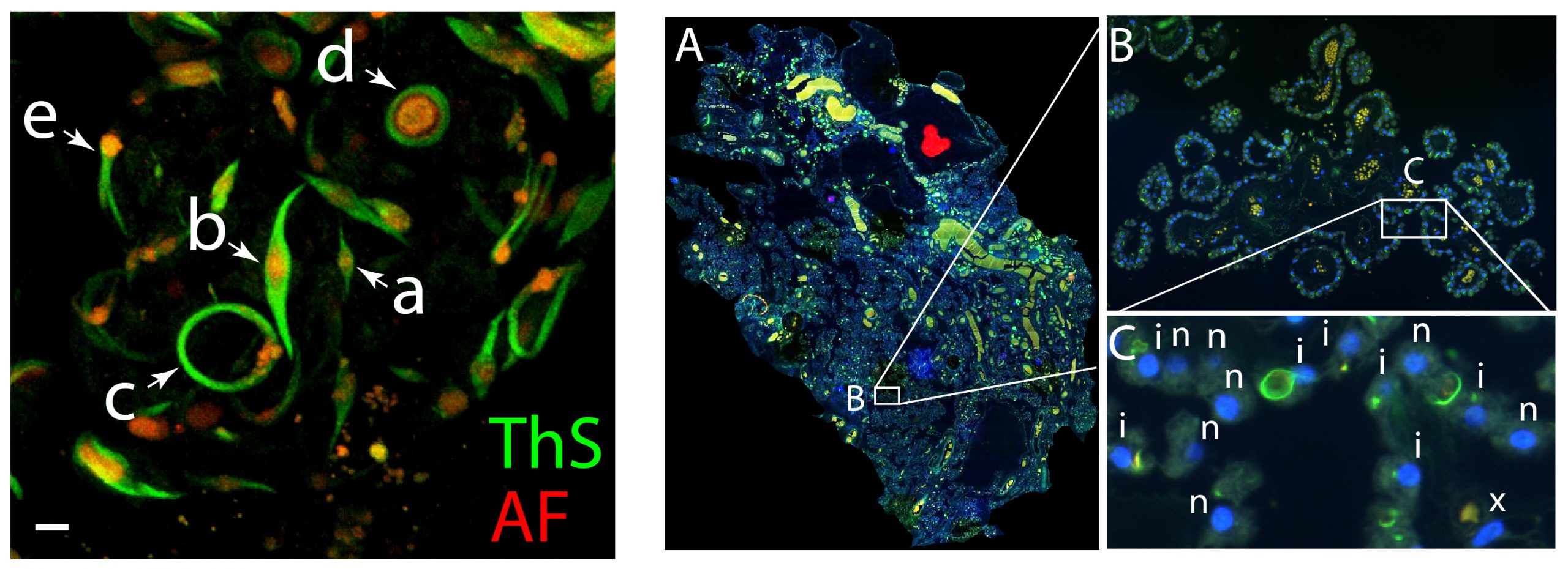
Although the choroid plexus (ChP) has been studied intermittently for over a century, more recent studies using modern techniques of staining, visualization, and rigorous quantification are quite scant. The older literature points to numerous human ChP features that accumulate normally with age and in an accelerated fashion in neurologic diseases. These features include fibrosis of the ChP stroma and three in CPECs – vacuoles of lipid/lipofuscin, mitochondrial accumulation (“oncocytic transformation”), and Biondi bodies. Biondi bodies are poorly characterized, lipid-associated amyloid aggregates of remarkably refined and varied structure that are relatively specific to CPECs and to humans. Using our tissue repository, we are studying these and other features in human ChP from different ventricles and the relationships of these pathologies to aging and diseases.
Biondi Bodies
For these mysterious inclusions, novel methods to visualize, quantify, and categorize Biondi bodies in 2-D and 3-D have been developed. Whole slide images scanned in the ADRC Neuropathology Core are used to count and map Biondi body distributions across large expanses of ChP, while their functional impacts on CPECs are also being explored. These data are then cross correlated with patient or participant metadata such as age, gender, ethnicity, diseases (e.g. Alzheimer’s and diabetes), and genotype (e.g. APOE). In collaboration with the High-End Mass Spectrometry Core, extracts of human postmortem tissues are being used to identify the amyloidogenic protein(s) that comprise Biondi bodies.
Artificial Intelligence
In addition to manual approaches with a team of undergraduate and volunteer researchers, we work with the Center of Artificial Intelligence in Diagnostic Medicine (CAIDM) on convolutional neural networks to identify and characterize human ChP pathologies. These AI approaches have already proven effective in identifying Biondi bodies in both 2-D and 3-D images, closely approaching the manual annotations.
