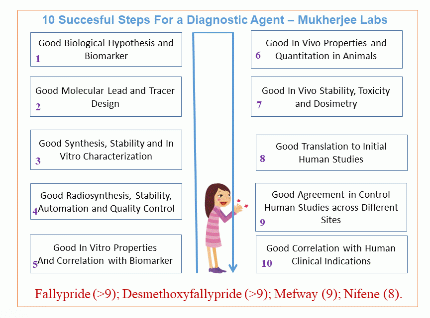
Molecular targeting of specific receptors, transporters, enzyme s and other biomarkers are carried out by identifying and/or designing small molecules that can be radiolabeled with radioisotopes for PET or SPECT imaging. Cellular and molecular markers (biomarkers) that may be potential indicators of disease progression are sought out for developing imaging probes. A dysregulation of the biomarker or a loss in the biomarker or an increase in the levels of the biomarker may be detectable by an in vivo imaging probe and assist in diagnosis of disease as well as treatment planning. Once a molecular target and/or biomarker is identified, the process of small molecule design and synthesis is undertaken. The graphic below shows the 10 step approach followed in our laboratories. This approach has been used successfully in our laboratories over the last three decades in optimizing imaging agents such as Fallypride and Desmethoxyfallypride for dopamine D2/D3 receptors, Mefway for serotonin 5HT1a receptors, Nifene for nicotinic receptors, all of which have now been taken to human studies.
Energy minimized structure of FALLYPRIDE which is used for PET imaging of dopamine D2 and D3 receptors. The yellow-green atom is fluorine-18 which is necessary for PET imaging.
Energy minimized structure of DESMETHOXYFALLYPRIDE, a dopamine D2/D3 receptor PET probe which acts faster than fallypride. The yellow-green atom is fluorine-18 which is necessary for PET imaging.
Energy minimized structure of NIFENE used for PET imaging of nicotinic a4b2 receptors. The yellow-green atom is fluorine-18 which is necessary for PET imaging.
