• Log In
  • Skip to primary navigation
  • Skip to content
  • Skip to primary sidebar

Mukherjee Labs Preclinical Imaging

Design and Development of Novel Imaging Methods for Early Diagnosis of Diseases

  • Mukherjee Labs – Preclinical Imaging
  • Research
    • Biomarkers Research and Development
    • Radiopharmaceutical Research
    • Pharmacology and Neuroscience
    • Autoradiography
    • Small Animal PET Imaging
    • Small Animal CT Imaging
    • Small Animal SPECT/CT
    • Human Brain & Whole Body PET Imaging
    • Bioluminescence and Fluorescence Optical Imaging
    • Data Sharing
  • People
    • Current Members
    • Former Members
  • Publications
    • Meeting Presentations
    • Schizophrenia
    • Alzheimer’s Disease
    • Parkinson’s Disease
    • Cancer
    • Diabetes & Obesity
  • News
    • Student Excellence in Research April 2025
    • Focused Ultrasound machine Feb 2025
    • UROP Symposium May 2024
    • Inveon Embedded PET/CT April 2024
    • Tau Imaging Research February 2024
    • New Hot Cells Installed May 2023
    • UROP UCI Symposium May 2023
    • Letter from US Congress; August 16, 2022
    • SNMMI 2022, JUNE 11-14
    • UROP Symposium May 2022
    • NRM 2021 December 14-16
    • Society for Neuroscience Mtg Nov 2021
    • WMIC, October 5-8, 2021
    • SNMMI Annual Meeting, June 12-15, 2021
    • Flotaza for Alzheimer’s disease
    • UROP UCI Symposium_ May 21, 2021
    • GE TracerLab FXN Installed 12/01/2020
    • New DCE Mini Hot Cell Installed 11/11/2020
    • World Molecular Imaging Conference (WMIC), 10/7/2020
    • New Inveon SPECT/CT Scanner Installed 9/23/2020
    • SNMMI Virtual Annual Meeting 2020, July 11-14
    • Metz Lectures, University of Chicago, Feb 4 and 5, 2020
    • 5th Int Symposium on Acetylcholine, Sept 27-29, 2019
    • SNMMI Annual Meeting, June 22-25, 2019
    • UROP UCI Symposium_ May 18, 2019
    • UROP UCI Symposium_ May 19, 2018
    • AALAS Imaging Workshop May 9, 2018
    • New Website
  • Support
  • Contact Us
You are here: Home / Mukherjee Lab – Preclinical Imaging / Research / Small Animal CT Imaging

Small Animal CT Imaging

The facility is equipped with a state of the art Inveon Multimodality (MM) CT  from Siemens, which has a resolution of up to 30 microns. The high resolution of the CT allows various applications for orthopedics, cardiology, oncology and others. The Inveon PET and CT are installed in tandem and can be used to carry out PET/CT imaging.


Contrast MicroCT imaging of the Spleen: A. No contrast agent injected; B. EXIA 160-XL was injected i.v. 30 min prior to microCT imaging of abdomen.  Spleen was clearly identified; C. Time course of CT contrast in the spleen.


Mouse CT Scans Lung Tumors. Representative CT scans of (A) control and (B) NNK-treated AJ mice, showing lung tumors (arrows) 10 months after NNK treatment. (C) Time-course changes of the volumes of lung tumor growth in the NNK-treated AJ mice (data are mean ± SD, p <0.05 for the different age groups). Baseline measures prior to NNK treatment indicate lung regions in the same animals were taken based on the contrast and may not necessarily reflect tumors

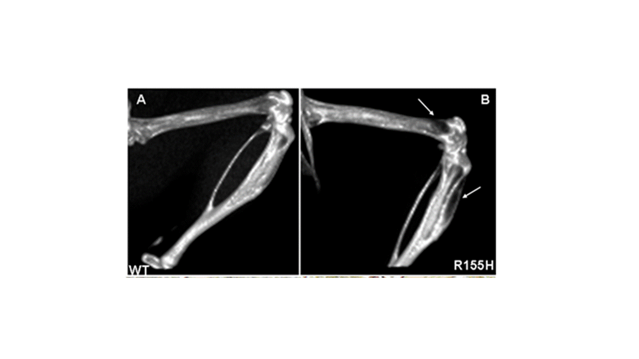
Bone MicroCT Images of Paget disease mice models:  (A-B) micro CT images showing sclerotic lesions at anterior tibia and posterior femur shown by white arrows in knock-in mice.


Primary Sidebar

Translational PET

Inveon PET

Inveon CT

Inveon SPECT/CT

ECAT HR+ PET

Molecular Imaging

Mukherjee Labs

Preclinical Imaging
Radiological Sciences
University of California Irvine
Irvine, CA 92697-5000
email: j.mukherjee@uci.edu
Tel: 949-824-2018

© 2025 UC Regents