Chadarevian et al. publish in Neuron

Therapeutic potential of human microglia transplantation in a chimeric model of CSF1R-related leukoencephalopathy

Microglia replacement strategies are increasingly being considered for the treatment of primary microgliopathies like adult-onset leukoencephalopathy with axonal spheroids and pigmented glia (ALSP). However, available mouse models fail to recapitulate the diverse neuropathologies and reduced microglia numbers observed in patients. In this study, we generated a xenotolerant mouse model lacking the fms-intronic regulatory element (FIRE) enhancer within Csf1r, which develops nearly all the hallmark pathologies associated with ALSP. Remarkably, transplantation of human induced pluripotent stem cell (iPSC)-derived microglial (iMG) progenitors restores a homeostatic microglial signature and prevents the development of axonal spheroids, white matter abnormalities, reactive astrocytosis, and brain calcifications. Furthermore, transplantation of CRISPR-corrected ALSP-patient-derived iMG reverses pre-existing spheroids, astrogliosis, and calcification pathologies. Together with the accompanying study by Munro and colleagues, our results demonstrate the utility of FIRE mice to model ALSP and provide compelling evidence that iMG transplantation could offer a promising new therapeutic strategy for ALSP and perhaps other microglia-associated neurological disorders.

Chadarevian et al. publish in Journal of Experimental Medicine

Engineering an inhibitor-resistant human CSF1R variant for microglia replacement

Hematopoietic stem cell transplantation (HSCT) can replace endogenous microglia with circulation-derived macrophages but has high mortality. To mitigate the risks of HSCT and expand the potential for microglia replacement, we engineered an inhibitor-resistant CSF1R that enables robust microglia replacement. A glycine to alanine substitution at position 795 of human CSF1R (G795A) confers resistance to multiple CSF1R inhibitors, including PLX3397 and PLX5622. Biochemical and cell-based assays show no discernable gain or loss of function. G795A- but not wildtype-CSF1R expressing macrophages efficiently engraft the brain of PLX3397-treated mice and persist after cessation of inhibitor treatment. To gauge translational potential, we CRISPR engineered human-induced pluripotent stem cell–derived microglia (iMG) to express G795A. Xenotransplantation studies demonstrate that G795A-iMG exhibit nearly identical gene expression to wildtype iMG, respond to inflammatory stimuli, and progressively expand in the presence of PLX3397, replacing endogenous microglia to fully occupy the brain. In sum, we engineered a human CSF1R variant that enables nontoxic, cell type, and tissue-specific replacement of microglia.

Shabestari et al publish in Cell Reports

Absence of microglia promotes diverse pathologies and early lethality in Alzheimer’s disease mice

Microglia are strongly implicated in the development and progression of Alzheimer’s disease (AD), yet their impact on pathology and lifespan remains unclear. Here we utilize a CSF1R hypomorphic mouse to generate a model of AD that genetically lacks microglia. The resulting microglial-deficient mice exhibit a profound shift from parenchymal amyloid plaques to cerebral amyloid angiopathy (CAA), which is accompanied by numerous transcriptional changes, greatly increased brain calcification and hemorrhages, and premature lethality. Remarkably, a single injection of wild-type microglia into adult mice repopulates the microglial niche and prevents each of these pathological changes. Taken together, these results indicate the protective functions of microglia in reducing CAA, blood-brain barrier dysfunction, and brain calcification. To further understand the clinical implications of these findings, human AD tissue and iPSC-microglia were examined, providing evidence that microglia phagocytose calcium crystals, and this process is impaired by loss of the AD risk gene, TREM2.

Jairaman and McQuade et al publish in eLife

TREM2 regulates purinergic receptor-mediated calcium signaling and motility in human iPSC-derived microglia

The membrane protein TREM2 (Triggering Receptor Expressed on Myeloid cells 2) regulates key microglial functions including phagocytosis and chemotaxis. Loss-of-function variants of TREM2 are associated with increased risk of Alzheimer’s disease (AD). Because abnormalities in Ca2+ signaling have been observed in several AD models, we investigated TREM2 regulation of Ca2+ signaling in human induced pluripotent stem cell-derived microglia (iPSC-microglia) with genetic deletion of TREM2. We found that iPSC-microglia lacking TREM2 (TREM2 KO) show exaggerated Ca2+ signals in response to purinergic agonists, such as ADP, that shape microglial injury responses. This ADP hypersensitivity, driven by increased expression of P2Y12 and P2Y13 receptors, results in greater release of Ca2+ from the endoplasmic reticulum (ER) stores, which triggers sustained Ca2+ influx through Orai channels and alters cell motility in TREM2 KO microglia. Using iPSC-microglia expressing the genetically encoded Ca2+ probe, Salsa6f, we found that cytosolic Ca2+ tunes motility to a greater extent in TREM2 KO microglia. Despite showing greater overall displacement, TREM2 KO microglia exhibit reduced directional chemotaxis along ADP gradients. Accordingly, the chemotactic defect in TREM2 KO microglia was rescued by reducing cytosolic Ca2+ using a P2Y12 receptor antagonist. Our results show that loss of TREM2 confers a defect in microglial Ca2+ response to purinergic signals, suggesting a window of Ca2+ signaling for optimal microglial motility.

Dr. Claes et al publish in Alzheimer’s and Dementia

The P522R protective variant of PLCG2 promotes the expression of antigen presentation genes by human microglia in an Alzheimer’s disease mouse model

The P522R variant of PLCG2, expressed by microglia, is associated with a reduced risk of Alzheimer’s disease (AD). Yet, the impact of this protective mutation on microglial responses to AD pathology remains unknown. Chimeric AD and wild-type mice were generated by transplanting PLCG2-P522R or isogenic wild-type human induced pluripotent stem cell microglia. At 7 months of age, single-cell and bulk RNA sequencing, and histological analyses were performed. The PLCG2-P522R variant induced a significant increase in microglial human leukocyte antigen (HLA) expression and the induction of antigen presentation, chemokine signaling, and T cell proliferation pathways. Examination of immune-intact AD mice further demonstrated that the PLCG2-P522R variant promotes the recruitment of CD8+ T cells to the brain. These data provide the first evidence that the PLCG2-P522R variant increases the capacity of microglia to recruit T cells and present antigens, promoting a microglial transcriptional state that has recently been shown to be reduced in AD patient brains.

Claes et al. publish in Molecular Neurodegeneration

Plaque-associated human microglia accumulate lipid droplets in a chimeric model of Alzheimer’s disease

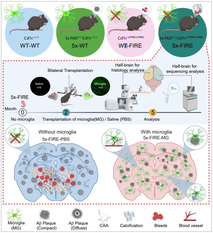
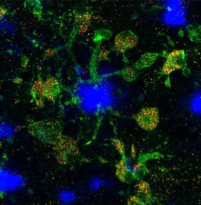
Background: Disease-associated microglia (DAMs), that surround beta-amyloid plaques, represent a transcriptionally-distinct microglial profile in Alzheimer’s disease (AD). Activation of DAMs is dependent on triggering receptor expressed on myeloid cells 2 (TREM2) in mouse models and the AD TREM2-R47H risk variant reduces microglial activation and plaque association in human carriers. Interestingly, TREM2 has also been identified as a microglial lipid-sensor, and recent data indicates lipid droplet accumulation in aged microglia, that is in turn associated with a dysfunctional proinflammatory phenotype. However, whether lipid droplets (LDs) are present in human microglia in AD and how the R47H mutation affects this remains unknown.

Methods: To determine the impact of the TREM2 R47H mutation on human microglial function in vivo, we transplanted wild-type and isogenic TREM2-R47H iPSC-derived microglial progenitors into our recently developed chimeric Alzheimer mouse model. At 7 months of age scRNA-seq and histological analyses were performed.

Results: Here we report that the transcriptome of human wild-type TREM2 and isogenic TREM2-R47H DAM xenografted microglia (xMGs), isolated from chimeric AD mice, closely resembles that of human atherosclerotic foam cells. In addition, much like foam cells, plaque-bound xMGs are highly enriched in lipid droplets. Somewhat surprisingly and in contrast to a recent in vitro study, TREM2-R47H mutant xMGs exhibit an overall reduction in the accumulation of lipid droplets in vivo. Notably, TREM2-R47H xMGs also show overall reduced reactivity to plaques, including diminished plaque-proximity, reduced CD9 expression, and lower secretion of plaque-associated APOE.

Conclusions: Altogether, these results indicate lipid droplet accumulation occurs in human DAM xMGs in AD, but is reduced in TREM2-R47H DAM xMGs, as it occurs secondary to TREM2-mediated changes in plaque proximity and reactivity.

McQuade et al. publish in Nature Communications

Gene expression and functional deficits underlie TREM2-knockout microglia responses in human models of Alzheimer’s disease

The discovery of TREM2 as a myeloid-specific Alzheimer’s disease (AD) risk gene has accelerated research into the role of microglia in AD. While TREM2 mouse models have provided critical insight, the normal and disease-associated functions of TREM2 in human microglia remain unclear. To examine this question, we profile microglia differentiated from isogenic, CRISPR-modified TREM2-knockout induced pluripotent stem cell (iPSC) lines. By combining transcriptomic and functional analyses with a chimeric AD mouse model, we find that TREM2 deletion reduces microglial survival, impairs phagocytosis of key substrates including APOE, and inhibits SDF-1α/CXCR4-mediated chemotaxis, culminating in an impaired response to beta-amyloid plaques in vivo. Single-cell sequencing of xenotransplanted human microglia further highlights a loss of disease-associated microglial (DAM) responses in human TREM2 knockout microglia that we validate by flow cytometry and immunohistochemistry. Taken together, these studies reveal both conserved and novel aspects of human TREM2 biology that likely play critical roles in the development and progression of AD.


Hasselmann and Blurton-Jones publish in Glia

Human iPSC-derived microglia: A growing toolset to study the brain’s innate immune cells

Recent advances in the generation of microglia from human induced pluripotent stem cells (iPSCs) have provided exciting new approaches to examine and decipher the biology of microglia. As these techniques continue to evolve to encompass more complex in situ and in vivo paradigms, so too have they begun to yield novel scientific insight into the genetics and function of human microglia. As such, researchers now have access to a toolset comprised of three unique “flavors” of iPSC-derived microglia: in vitro microglia (iMGs), organoid microglia (oMGs), and xenotransplanted microglia (xMGs). The goal of this review is to discuss the variety of research applications that each of these techniques enables and to highlight recent discoveries that these methods have begun to uncover. By presenting the research paradigms in which each model has been successful, as well as the key benefits and limitations of each approach, it is our hope that this review will help interested researchers to incorporate these techniques into their studies, collectively advancing our understanding of human microglia biology.

Hasselmann and Coburn et al. publish in Neuron

Development of a Chimeric Model to Study and Manipulate Human Microglia In Vivo

iPSC-derived microglia offer a powerful tool to study microglial homeostasis and disease-associated inflammatory responses. Yet, microglia are highly sensitive to their environment, exhibiting transcriptomic deficiencies when kept in isolation from the brain. Furthermore, species-specific genetic variations demonstrate that rodent microglia fail to fully recapitulate the human condition. To address this, we developed an approach to study human microglia within a surrogate brain environment. Transplantation of iPSC-derived hematopoietic-progenitors into the postnatal brain of humanized, immune-deficient mice results in context-dependent differentiation into microglia and other CNS macrophages, acquisition of an ex vivo human microglial gene signature, and responsiveness to both acute and chronic insults. Most notably, transplanted microglia exhibit robust transcriptional responses to Aβ-plaques that only partially overlap with that of murine microglia, revealing new, human-specific Aβ-responsive genes. We therefore have demonstrated that this chimeric model provides a powerful new system to examine the in vivo function of patient-derived and genetically modified microglia.

McQuade et al. publish in Molecular Neurodegeneration

Development and validation of a simplified method to generate human microglia from pluripotent stem cells

Microglia, the principle immune cells of the brain, play important roles in neuronal development, homeostatic function and neurodegenerative disease. Recent genetic studies have further highlighted the importance of microglia in neurodegeneration with the identification of disease risk polymorphisms in many microglial genes. To better understand the role of these genes in microglial biology and disease, we, and others, have developed methods to differentiate microglia from human induced pluripotent stem cells (iPSCs). While the development of these methods has begun to enable important new studies of microglial biology, labs with little prior stem cell experience have sometimes found it challenging to adopt these complex protocols. Therefore, we have now developed a greatly simplified approach to generate large numbers of highly pure human microglia.