New Study Exposes Hidden Impact of Childhood Trauma on the Brain—Revolutionizing Mental Health Care.

A groundbreaking study has revealed the powerful impact of adverse childhood experiences (ACEs) on brain function, offering critical insights into how early trauma shapes mental health in adulthood. Published today in Frontiers in Psychiatry, this is the largest brain-imaging study ever conducted on ACEs.

The study involved over 7,000 participants and used brain SPECT imaging and clinical evaluations to map how childhood trauma affects brain activity. Results show that higher ACE scores are closely linked to an increased risk of psychiatric disorders, including PTSD, anxiety, depression, and substance abuse.

Crucial findings revealed abnormal activity in areas of the brain associated with emotional pain and decision-making, such as the anterior cingulate and thalamus. These areas, responsible for processing both physical and emotional suffering, were overactive in individuals with high ACE scores, explaining the link between childhood trauma and conditions like fibromyalgia and chronic migraines.

New Landmark Study Reveals Brain Blood Flow as Key Biomarker in Depression

A major international study, published this week in JAMA Psychiatry, has identified a powerful new brain-based biomarker for Major Depressive Disorder (MDD), marking a potential turning point in how depression is diagnosed and treated. The study used regional cerebral blood flow (RCBF) data measured by brain SPECT imaging to validate a reproducible, regionally specific pattern of hypoperfusion in the brains of individuals suffering from depression.

This landmark research demonstrates that functional brain changes—specifically decreased blood flow—are more accurate markers of depression than structural brain changes like cortical thinning, which have historically shown weak and inconsistent associations.

The study examined more than 15,000 participants across four major databases, including the UK Biobank, ENIGMA Consortium, Amish Connectome Project, and Amen Clinics Inc. Notably, the blood flow patterns from SPECT scans aligned strongly with novel MRI-based measures of brain function (ReHo), confirming that decreased activity in the cingulate, prefrontal, and temporal lobes is a hallmark of depression.

Even more importantly, the study found that these blood flow deficits were strongly correlated with the severity of depressive symptoms, opening the door for precision diagnostics and individualized treatment plans based on imaging biomarkers.

Chronic Negativity Linked to Poor Memory, Depression, and Brain Dysfunction

A groundbreaking new study accepted for publication in Depression and Anxiety reveals that individuals with high levels of conscious negativity bias—where the mind fixates on potential dangers or worst-case scenarios—show significant dysfunction in brain regions tied to emotional regulation and cognitive control.

Researchers analyzed brain scans and cognitive data from 1,984 patients with anxiety disorders, using advanced SPECT imaging and the Total Brain assessment platform. The findings were striking: higher negativity bias was associated with decreased activity in the frontal and temporal lobes, the insula, and parietal regions—all areas involved in focus, decision-making, and emotional stability. In contrast, increased activity was observed in specific cerebellar lobules, linked to motor tension and emotional overactivation.

Patients with elevated negativity bias also reported significantly higher levels of anxiety, depression, suicidal ideation, emotional instability, and poor memory and stress regulation.

The study underscores the importance of addressing negativity bias in therapy—not just for emotional relief but also for improving brain health and cognitive performance. Researchers recommend positivity bias training, including simple daily practices like:

  • Starting the day by saying: “Today is going to be a great day,”
  • Actively looking for the micro-moments of happiness during the day,
  • And ending each day by reflecting on: “What went well today?”

New Publication: Joint-label fusion brain atlases for dementia research in Down syndrome

Research suggests a link between Alzheimer’s Disease in Down Syndrome (DS) and the overproduction of amyloid plaques. Using Positron Emission Tomography (PET) we can assess the in-vivo regional amyloid load using several available ligands. To measure amyloid distributions in specific brain regions, a brain atlas is used. A popular method of creating a brain atlas is to segment a participant’s structural Magnetic Resonance Imaging (MRI) scan. Acquiring an MRI is often challenging in intellectually-imparied populations because of contraindications or data exclusion due to significant motion artifacts or incomplete sequences related to general discomfort. When an MRI cannot be acquired, it is typically replaced with a standardized brain atlas derived from neurotypical populations (i.e. healthy individuals without DS) which may be inappropriate for use in DS. In this project, we create a series of disease and diagnosis-specific (cognitively stable (CS-DS), mild cognitive impairment (MCI-DS), and dementia (DEM-DS)) probabilistic group atlases of participants with DS and evaluate their accuracy of quantifying regional amyloid load compared to the individually-based MRI segmentations. Further, we compare the diagnostic-specific atlases with a probabilistic atlas constructed from similar-aged cognitively-stable neurotypical participants. We hypothesized that regional PET signals will best match the individually-based MRI segmentations by using DS group atlases that aligns with a participant’s disorder and disease status (e.g. DS and MCI-DS). Our results vary by brain region but generally show that using a disorder-specific atlas in DS better matches the individually-based MRI segmentations than using an atlas constructed from cognitively-stable neurotypical participants. We found no additional benefit of using diagnose-specific atlases matching disease status. All atlases are made publicly available for the research community.View Publication

New Publication: Linear Regression Tool for NIDM Documents

Congratulations to Ashmita Kumar on her first scientific publication!

Abstract

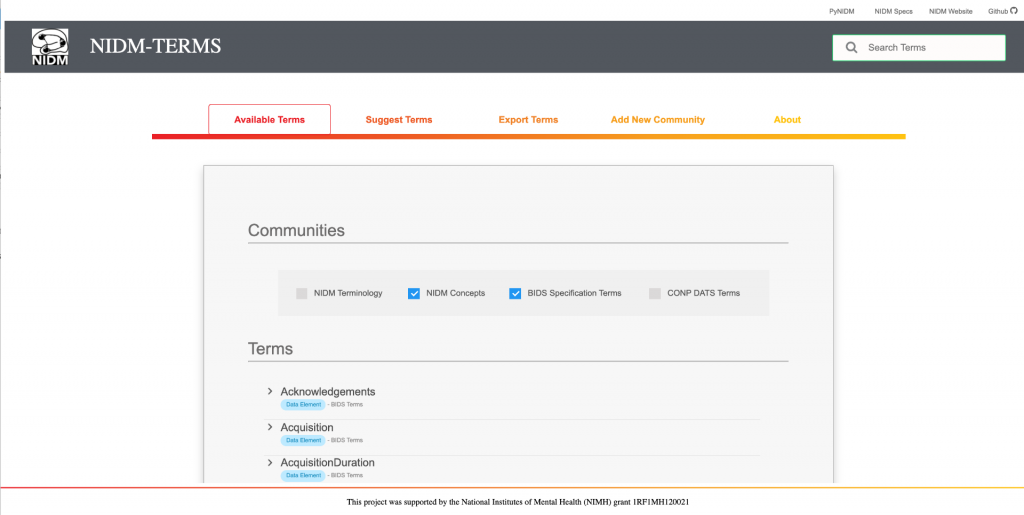
The Neuroimaging Data Model (NIDM) is a series of specifications for describing all aspects of the neuroimaging data lifecycle from raw data to analyses and provenance. NIDM uses community-driven terminologies along with unambiguous data dictionaries within a Resource Description Framework (RDF) document to describe data and metadata for integration and query. Data from different studies, using locally defined variable names, can be retrieved by linking them to higher-order concepts from established ontologies and terminologies. Through these capabilities, NIDM documents are expected to improve reproducibility and facilitate data discovery and reuse. PyNIDM is a Python toolbox supporting the creation, manipulation, and querying of NIDM documents. Using the query tools available in PyNIDM, users are able interrogate datasets to find studies that have collected variables measuring similar phenotypic properties. This, in turn, facilitates the transformation and combination of data across multiple studies.
Full Paper: https://f1000research.com/articles/11-228/v1

New Publication: Alzheimer‐related altered white matter microstructural integrity in Down syndrome: A model for sporadic AD?

Introduction

Virtually all adults with Down syndrome (DS) develop Alzheimer’s disease (AD)‐associated neuropathology by the age of 40, with risk for dementia increasing from the early 50s. White matter (WM) pathology has been reported in sporadic AD, including early demyelination, microglial activation, loss of oligodendrocytes and reactive astrocytes but has not been extensively studied in the at‐risk DS population.

Methods

Fifty‐six adults with DS (35 cognitively stable adults, 11 with mild cognitive impairment, 10 with dementia) underwent diffusion‐weighted magnetic resonance imaging (MRI), amyloid imaging, and had assessments of cognition and functional abilities using tasks appropriate for persons with intellectual disability.

Results

Early changes in late‐myelinating and relative sparing of early‐myelinating pathways, consistent with the retrogenesis model proposed for sporadic AD, were associated with AD‐related cognitive deficits and with regional amyloid deposition.

Discussion

Our findings suggest that quantification of WM changes in DS could provide a promising and clinically relevant biomarker for AD clinical onset and progression.

https://alz-journals.onlinelibrary.wiley.com/doi/10.1002/dad2.12040

NIH Awards Over $100 Million to Examine Biomarkers of Alzheimer’s Disease in Adults with Down Syndrome

he Alzheimer’s Biomarkers Consortium – Down Syndrome (ABC-DS), a multi-institution research team, co-led by members from the University of California, Irvine, has been awarded an unprecedented five-year, $109 million grant by the National Institutes of Health (NIH), to expand research on the biomarkers of Alzheimer’s disease in adults with Down syndrome.

https://som.uci.edu/news_releases/ABC-DS-grant-awarded.asp?fbclid=IwAR3fuTF3dXAfjKezO3CaNtcEBbd31wIOPEdXRRKLYtbh8lJDwRPDeCHX7_o