
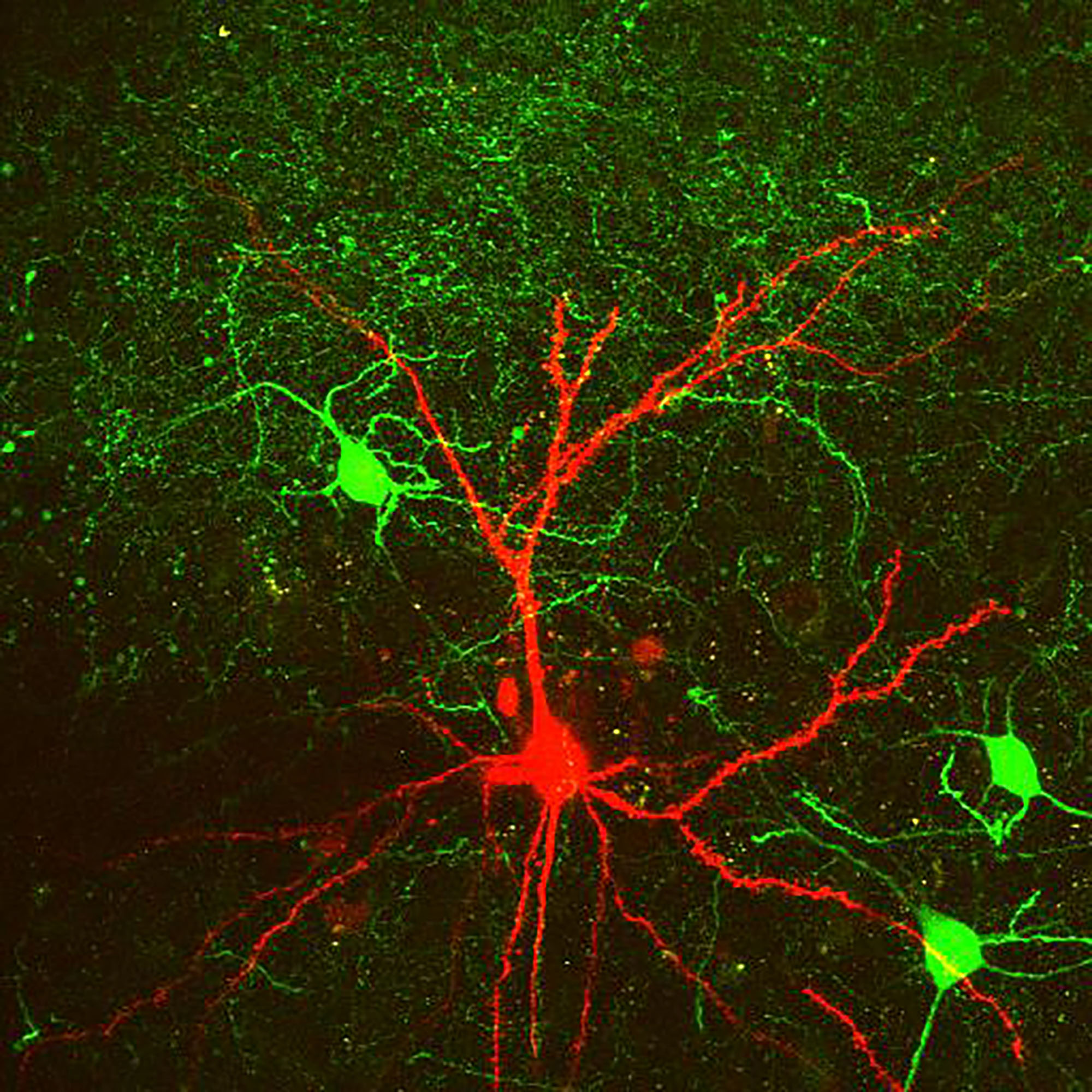
Patch-clamp recording
Today the patch-clamp technique is considered to be the gold standard method in neuroscience allowing us to record the electrical activity of neurons with high sensitivity and temporal precision. In the Lur lab we use whole-cell recordings from fluorescently identified neurons to measure the spatial and temporal integration of various synaptic inputs and to map the connectivity of local circuits.

Optical interrogation of neural circuits
To gain a deeper understanding of how the brain works, we aim to establish a causal relationship between the activity of a given neural circuit and its downstream consequences (e.g. firing of the target neurons or the behavior of an animal). This cannot be achieved by only observing the brain; to establish causality we need to manipulate neuronal activity in a controlled manner. To this end, we use targeted expression of optogenetic tools that allow us to control the output of a desired cell population. We then measure the effect of these manipulations on the circuit’s output in vitro or in vivo.
Multi-photon imaging
The introduction of multi-photon imaging revolutionized neuroscience. State of the art techniques today allow us to use 2-photon microscopy to record biochemical signals in the smallest neuronal compartments (dendritic spines), map synaptic inputs on the dendritic arbour of multimodal neurons, or monitor the activity of hundreds of neurons simultaneously in response to an environmental stimulus.